
The Degeneracy of the Genetic Code
The Degeneracy of the Genetic Code
Author: Shenzhou
Reviewed by: Shenzhou
During translation, the genetic code maps a nucleotide sequence to an amino acid sequence, while tRNAs physically carry out that mapping. The code is therefore the essential link between nucleic acids and proteins.
Early mathematical analyses led scientists to conclude that codons had to contain three bases to encode all 20 amino acids. Crick later inserted or deleted different numbers of bases in bacteriophages and compared the resulting phenotypes. His experiments supported the hypothesis that the genetic code is read in nucleotide triplets.
At about the same time, Nirenberg and Matthaei used synthetic mRNA and an in vitro translation system to match several simple codons, including UUU, CCC, and AAA, to their amino acids. Soon afterward, Nirenberg continued decoding the genetic code with a nitrocellulose membrane that retained complexes of ribosomes, mRNA, and aminoacyl-tRNA.
The researchers prepared 20 in vitro systems, each containing ribosomes and all 20 amino acids. In each system, a different amino acid was labeled with carbon-14 (C14). They then added a synthetic trinucleotide RNA and filtered the reaction mixture through nitrocellulose. In principle, only a complete ribosome–amino acid–tRNA–mRNA complex would remain on the membrane and produce a strong radioactive signal. By repeating the test, they could determine which trinucleotide corresponded to each amino acid. Scientists eventually used this method to decode all 61 non-stop codons.
Stop codons encode no amino acid, so this method could not reveal them. In 1965, Garen found another approach: he inferred stop-codon sequences by studying reversion in E. coli nonsense mutants.
An amber mutant of the E. coli alkaline phosphatase gene had arisen when a tryptophan codon (UGG) mutated into a stop codon. Garen obtained many revertants, identified the new amino acid at that site in each one, and analyzed its codon: Ser (UCG), Leu (UUG), Tyr (UAC), Lys (AAG), Gln (CAG), or Glu (GAG). Comparing these sequences showed that the stop codon derived from Trp (UGG) was UAG, identifying the first stop codon. In 1967, Brenner and Crick used the same approach to identify the last one. The three stop codons were named for the nonsense mutations used in this work: UAG, amber; UAA, ochre; and UGA, opal.
Once the amino-acid codons had been identified, one feature stood out: there are many more codons than amino acids. This redundancy is called the degeneracy of the genetic code. Leu, for example, is encoded by four codons: CUU, CUC, CUA, and CUG. Across many such groups, the first two bases remain fixed while the third varies. The third position therefore often plays a smaller role in determining the amino acid, a pattern that contributes to the code's degeneracy.
A tRNA's anticodon sits in a loop, so its three bases follow a curve, while the mRNA codon lies in a straight line. The two therefore do not align with perfectly regular geometry. The structure of tRNA also gives the first anticodon base considerable freedom. As a result, that base can pair with the codon's third base without following the usual complementary base-pairing rules exactly. Chemical modification at this anticodon position permits still more combinations. This “wobble” pairing allows just 32 tRNAs to read 61 codons, reducing the number of genes needed to encode tRNAs.
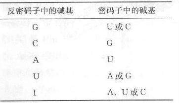
This degeneracy gives genes a substantial margin for error and reduces the harm caused by mutation. A nucleotide change at some sites may leave the resulting protein, and therefore the organism's traits, unchanged. This helps preserve genetic stability across generations. The organization of the code offers another layer of protection as well.
If the third codon position often matters less, what roles do the first two play? Part of the answer lies in the physicochemical properties of amino acids.
When codons are compared with amino-acid polarity, codons that share the same second base often encode amino acids with similar physical or chemical properties. In general, C in the second position is associated with nonpolar amino acids or those with uncharged side chains; U often corresponds to strongly hydrophobic amino acids; and A or G generally corresponds to hydrophilic ones. If a mutation leaves the second codon position unchanged, the substituted amino acid may therefore retain properties similar to those of the original. This further limits the damage a mutation can cause.
All articles in this blog are licensed under CC BY-NC-SA 4.0 unless stating additionally.
 爱发电(点击图片跳转)