
Protein Translation
Protein Translation
Author: Shenzhou
Reviewed by: Shenzhou and Myelin
Proteins are the main functional molecules that sustain the activities of life, and the process by which cells synthesize them is called translation. During translation, mRNA serves as the template. With the help of the ribosome, tRNA interprets the nucleotide sequence in the mRNA as an amino acid sequence, producing a polypeptide chain. The principal participants are tRNA, rRNA, and mRNA. tRNA carries amino acids to the ribosome. rRNA joins with several ribosomal proteins to form the ribosome, the site of polypeptide synthesis. mRNA carries the codons that provide the template for translation.
mRNA
Jacob and Monod proposed the concept of messenger RNA in 1961. Because proteins are synthesized in the cytoplasm rather than in the nucleus, they reasoned that some substance must carry genetic information out of the nucleus. To demonstrate the existence of such a molecule, they designed several experiments involving the regulation of lactose metabolism in E. coli, bacterial conjugation, and density-gradient centrifugation.
Their experiments on the regulation of lactose metabolism in E. coli first showed that β-galactosidase is an “inducible enzyme,” expressed only when lactose is present. Bacterial conjugation experiments then showed that the information directing the synthesis of this enzyme was not carried directly by rRNA, but by another, short-lived substance. Finally, density-gradient centrifugation confirmed the existence of mRNA, distinguished it from rRNA, and showed that rRNA is a stable nucleic-acid molecule. In other words, rRNA reads the code during translation, while mRNA determines which protein is synthesized.
In prokaryotes, mRNA is transcribed directly from DNA in the cytoplasm, and translation begins while transcription is still under way. Prokaryotic mRNA is also short-lived, generally lasting only a few minutes. Eukaryotic mRNA is synthesized in the nucleus and spliced into mature mRNA before entering the cytoplasm, where it serves as the template for protein translation. Eukaryotic mRNA is more stable than its prokaryotic counterpart and can persist for several hours.
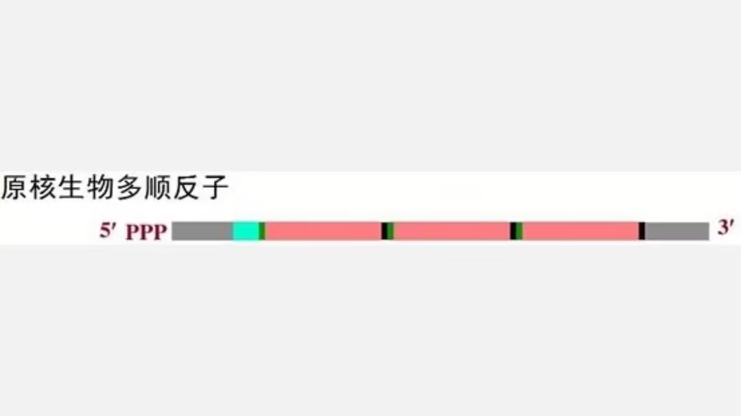
In prokaryotes, each cistron (p1) has its own ribosome-binding site upstream of the start codon: the -AGGAGGU- sequence, known as the Shine–Dalgarno sequence (SD sequence). A single prokaryotic mRNA can therefore direct the translation of several proteins.
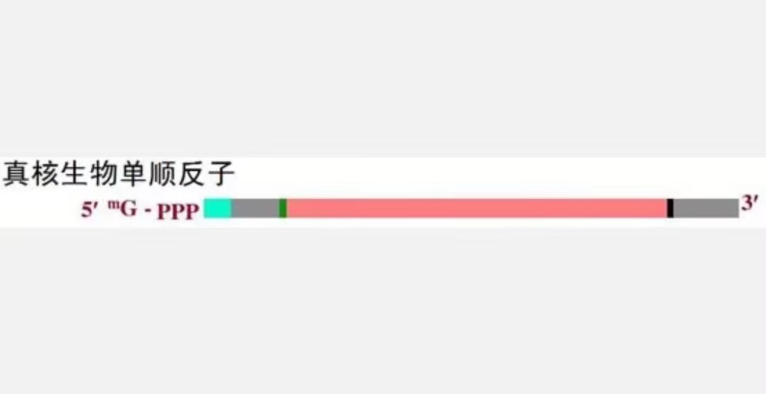
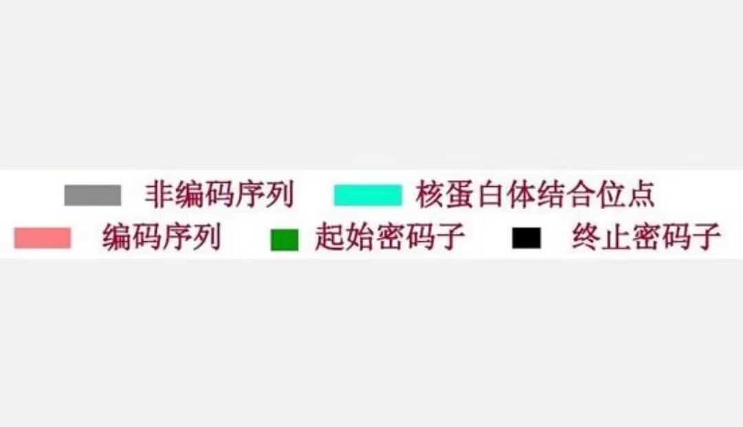
In eukaryotes (p2), by contrast, only the first AUG in an mRNA has an upstream CCACC signal sequence that the ribosome can recognize as it scans the transcript. A eukaryotic transcript can therefore contain only one cistron.
tRNA
During protein translation, tRNA acts as the adaptor between mRNA and amino acids. It ensures that amino acids are added to the polypeptide in the order specified by the codons in the mRNA.
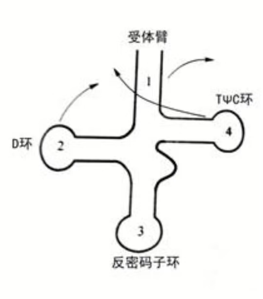
tRNA structure varies little among organisms and is highly conserved. With a few exceptions, including tRNAs in vertebrate mitochondria, all tRNAs have a cloverleaf secondary structure that supports their function. The following structural features allow tRNA to perform its various roles:
- A conserved CCU sequence at the 3’ end, which forms the amino-acid acceptor arm
- The D loop, which binds the corresponding aminoacyl-tRNA synthetase so that the matching amino acid can be loaded
- The anticodon loop, which contains the anticodon at positions 34, 35, and 36 and pairs with a codon during translation, allowing the codon to be recognized
- The TΨC loop, the principal ribosome-binding site, which helps form a stable translation complex
The secondary structure is shown below:
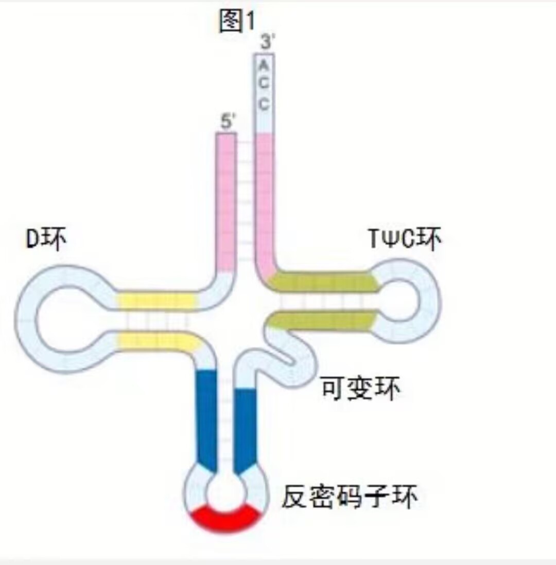
The cloverleaf represents only tRNA's secondary structure. Its three-dimensional structure resembles an inverted “L.” As this inverted-L structure forms, bases in two of the tRNA's loops, the D loop and the TΨC loop, pair as the molecule folds. The two diagrams below illustrate this folding process.
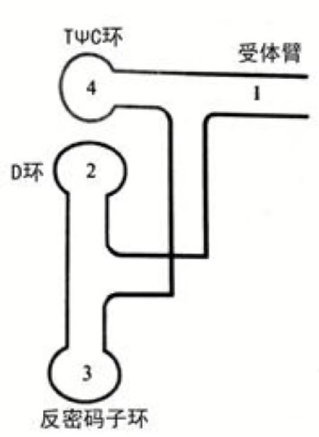
Continuous base pairing within tRNA produces base-stacking interactions that help stabilize its three-dimensional structure.
rRNA
The ribosome is the principal site of protein synthesis. This structurally complex ribonucleoprotein complex is abundant within the cell. E. coli contains roughly 20,000 ribosomes, and the average eukaryotic cell contains even more. Ribosomes can remain free in the cytoplasm; in eukaryotic cells, they can also attach to the rough endoplasmic reticulum.
Prokaryotic ribosomes are smaller, while eukaryotic ribosomes are somewhat larger, but both consist of two unequal subunits made up of several rRNAs and proteins. The E. coli ribosome is a 70S ribosome composed of a 30S small subunit, which contains 16S rRNA and 21 proteins, and a 50S large subunit, which contains 23S and 5S rRNA and 34 proteins. The eukaryotic 80S ribosome consists of a 40S small subunit, containing 33 proteins and 18S rRNA, and a 60S large subunit, containing 50 proteins and 28S, 5.8S, and 5S rRNA.
The large ribosomal subunit in both eukaryotes and prokaryotes contains 5S rRNA. By forming hydrogen-bonded base pairs with the TΨC loop of tRNA, this rRNA helps stabilize aminoacyl-tRNA binding to the ribosome.
Prokaryotic 16S rRNA and eukaryotic 18S rRNA are highly similar, though their structures differ somewhat because they recognize start codons by different mechanisms.
A complete ribosome has eight important structural domains and functional sites (p7). More broadly, however, it performs two main functions. First, it coordinates tRNA, mRNA, and the growing protein, keeping all three correctly positioned relative to one another during synthesis. Second, it catalyzes several key reactions in translation, allowing the process to continue smoothly through termination.
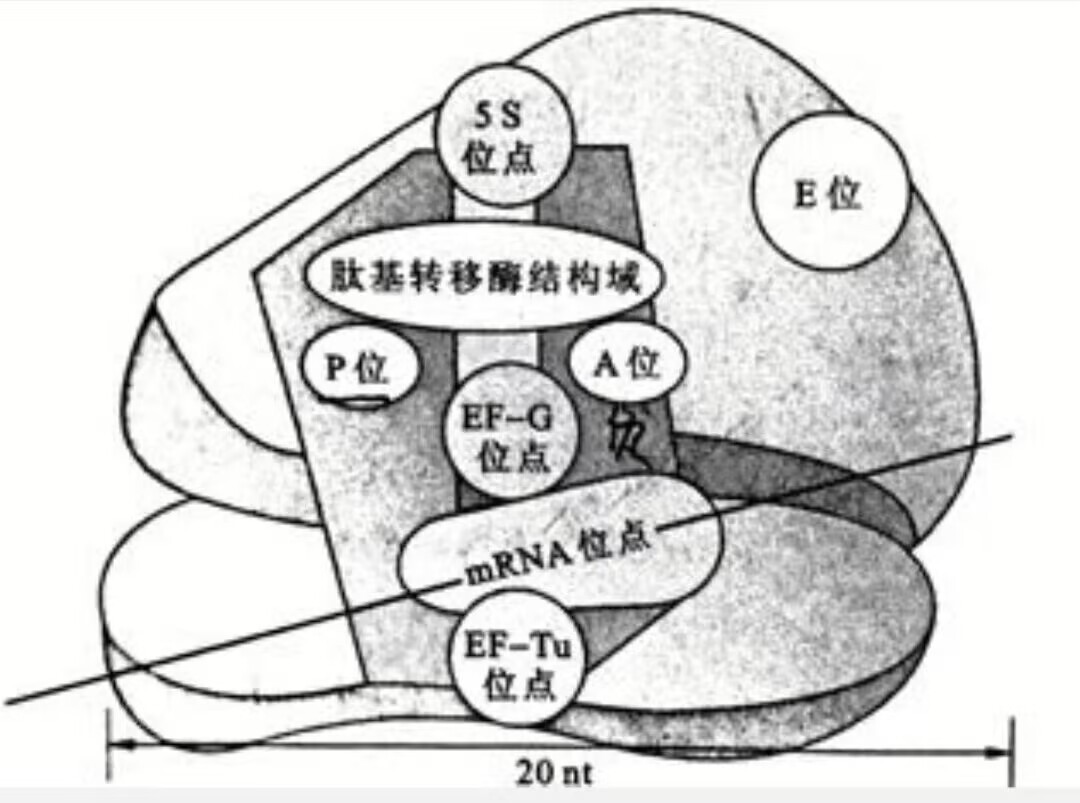
When translation begins, the large and small subunits temporarily assemble into a complete ribosome. Once synthesis is finished, the ribosome dissociates, and the two subunits return to the cytoplasm for the next round of translation.
All articles in this blog are licensed under CC BY-NC-SA 4.0 unless stating additionally.
 爱发电(点击图片跳转)