Fluorescent Proteins: A Brief History and How They Work
Author: Polar Glacier
Reviewed by: Weiming
Abstract
Green fluorescent protein (GFP), discovered and isolated from the jellyfish Aequorea victoria in 1962, and the red fluorescent protein drFP583, discovered in 1999 in corals of the genus Discosoma, have become widely used tools throughout biological research. They are nontoxic, emit strong fluorescence when excited by specific wavelengths of light, have low molecular weights, are easy to incorporate into vectors, and can be expressed in many organisms. These properties have made fluorescent proteins useful across a broad range of fields, and researchers now have many varieties to choose from.
History of Fluorescent-Protein Research
Green fluorescent protein was the first fluorescent protein discovered. In 1962, Japanese scientist Osamu Shimomura isolated it from the jellyfish Aequorea victoria while extracting aequorin. GFP looked green in sunlight, bright yellow under tungsten light, and vivid green under ultraviolet light. Through biochemical and spectroscopic studies, Shimomura began to unravel its fluorescence mechanism and identify its chromophore[1]. He could not have known that the discovery would transform modern biotechnology three decades later. Before long, he set GFP aside and moved on to other areas of research.
Between 1985 and 1992, Douglas Prasher determined GFP's gene and protein sequences and then cloned the gene in 1992. Martin Chalfie of Columbia University was the first to put GFP to work in biological research. In 1994, he introduced GFP cDNA into model organisms, including nematodes, and successfully expressed it, revealing the protein's enormous potential as a reporter. That same year, Roger Tsien's team elucidated GFP's fluorescence mechanism and engineered mutants with brighter fluorescence, a single absorption peak, and more efficient folding, including GFP-S65T[2].
In 1999, researchers reported a second type of fluorescent protein: the red fluorescent protein drFP583. Matz and his colleagues purified this tetrameric protein from Pacific corals of the genus Discosoma; under ultraviolet light, it fluoresces red. Its longer emission wavelength, high sensitivity, and strong signal-to-noise ratio made it a valuable complement to GFP in living organisms. Oligomerization, slow maturation, and cellular toxicity, however, limited the use of drFP583 itself. Researchers more often use E57-NA, an engineered mutant produced by Clontech and sold as DsRed, which has lower cytotoxicity, less tendency to oligomerize, and faster maturation. Continued improvements to DsRed have expanded its uses. Together, DsRed and GFP offer researchers in molecular biology, cell biology, and related fields a fast new way to detect biological processes[3].
How Fluorescent Proteins Work
Fluorescent-Protein Structure
Structure of Green Fluorescent Protein
The cDNA sequence reveals the primary structure of wild-type GFP: 238 amino-acid residues with a molecular mass of approximately 27 kDa[2]. X-ray diffraction analysis of GFP crystals[4] revealed a cylindrical structure about 4 nm high and 3 nm in diameter.
Eleven β-strands wrap around one another to form the wall of the barrel. They pack tightly together and are linked by regular networks of hydrogen bonds. A short α-helix caps one end, creating a densely packed structure with a single opening: the β-barrel.
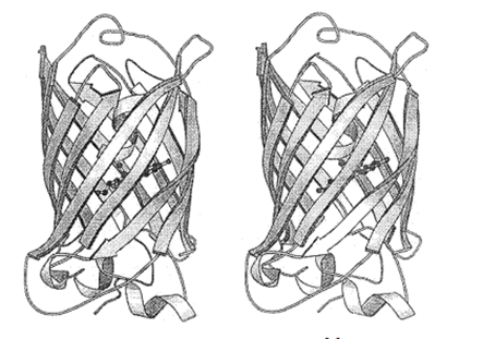 Figure 1. Secondary structure of GFP[4]
 The α-helix extends inward along the central axis and serves as a scaffold for the chromophore. At the center of the protein, the surrounding β-sheets protect the chromophore. This architecture makes GFP highly stable and helps the chromophore withstand heat, acids, bases, denaturants, and other stresses[5]. The compact structure also prevents enzymes and cofactors from reaching the interior to catalyze chromophore formation, so the chromophore must form autocatalytically.
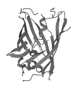 Figure 2. Structural relationship between the GFP chromophore and the β-barrel[6]
 The GFP chromophore forms from the Ser–Tyr–Gly tripeptide at residues 65–67. Autocatalytic cyclization produces 4-(p-hydroxybenzylidene)imidazolin-5-one, the structure at the heart of the chromophore.
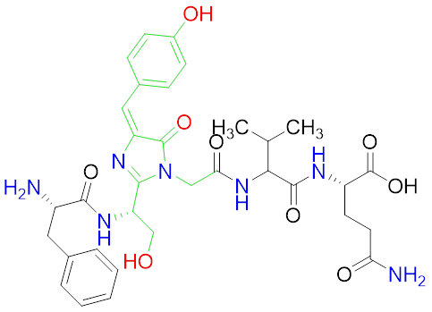 Figure 3. Structure of the GFP chromophore, with the remaining amino-acid residues omitted
 Chromophore formation[7] is an autocatalytic process that requires neither an enzyme nor a cofactor. Ser65 and Gly67 first cyclize rapidly to form an imidazolin-5-one intermediate. O2 then slowly oxidizes the Tyr66 side chain, creating a double bond. This step often takes several hours. Gly67 is indispensable; no other amino acid can take its place. The reaction is also temperature-sensitive, and its yield falls above 30 ℃, perhaps because the jellyfish naturally lives in colder water. Once formed, however, the chromophore is thermally stable.
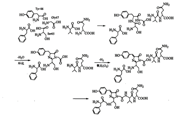 Figure 4. Formation of the GFP chromophore[7]
 Experiments have shown[7] that residues 2–232 of GFP are needed to maintain its fluorescence. Removing more than seven residues from the C terminus or more than two from the N terminus eliminates fluorescence altogether. The final seven residues are disordered and do not contribute to the protein's defined structure, so residues can be removed from or added to this region without affecting overall function. Residues at the N terminus help form the “cap” at one end of the β-barrel. This indispensable structure protects the chromophore, although extending the peptide chain at the N terminus does not disrupt the protein. In some GFP fusion proteins, removing several C-terminal residues does not eliminate fluorescence but can still alter the spectrum[5].
GFP can form dimers in crystals or in solutions with an ionic strength below 100 mmol/L. Under natural conditions, dimerization promotes energy transfer between GFP and the jellyfish photoprotein.
Structure of Red Fluorescent Protein (DsRed)
DsRed consists of 225 amino-acid residues and has a molecular mass of approximately 25.9 kDa. Although this coral protein shares only about 23% primary-sequence identity with GFP from Aequorea victoria, the two proteins have highly similar three-dimensional structures. Like GFP, DsRed forms a β-barrel.
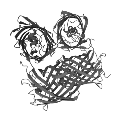 Figure 5. Secondary structure of DsRed, shown as a tetramer[8]
 The DsRed chromophore forms from Gln66–Tyr67–Gly68 and closely resembles the GFP chromophore. Early in its maturation in vitro, it has excitation and emission wavelengths of 475 nm and 499 nm, respectively, and therefore fluoresces green. The green fluorescence peaks after about 7 hours and then begins to fade. After 2 days, it disappears completely, and red fluorescence slowly emerges. The evidence suggests that DsRed first forms an intermediate almost identical to the GFP chromophore, then undergoes further oxidation. Unlike its GFP counterpart, the mature DsRed chromophore contains an imine group[9]. The imine double bond extends the conjugated system, shifting emission toward longer wavelengths.
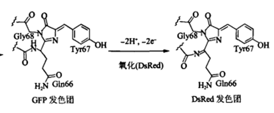 Figure 6. Formation of the DsRed chromophore passes through a GFP-like chromophore intermediate[9]
 Crystallographic analysis shows that DsRed normally exists as a tetramer. Experiments also indicate that its chromophore can form and function only when the protein oligomerizes. Denaturation studies using alkaline conditions, acids, and guanidine suggest that the chromophore forms through several folding steps and that oligomerization plays an important part in its maturation[3].
Mechanism of Fluorescence
The fluorescent proteins discovered so far all work in broadly similar ways. Because GFP was the first to be found and has been studied most extensively, this section focuses on its fluorescence mechanism.
In Aequorea victoria, GFP works in tandem with another light-producing protein, aequorin.
Aequorin contains coelenterazine as a prosthetic group and emits blue light at 460–470 nm in the presence of calcium ions.
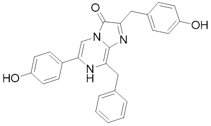 Figure 7. Coelenterazine
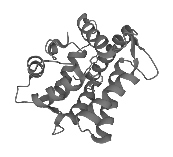 Figure 8. Aequorin[10]
 In the presence of calcium ions, oxygen oxidizes aequorin and the protein emits blue light. Förster resonance energy transfer carries that energy to the GFP chromophore, which then emits its characteristic green fluorescence at 509 nm.
GFP fluorescence is generally understood to involve excited-state proton transfer (ESPT)[11]. The key is the phenolic group of Tyr66 in the chromophore. Because phenol is weakly acidic, the chromophore can switch between neutral and ionic forms. Under ordinary conditions, the neutral form predominates at a ratio of approximately 6:1.
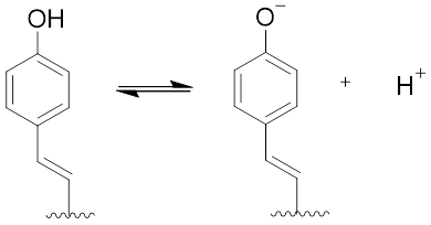 Figure 9. Interconversion between the neutral and ionic forms of the chromophore's phenolic group
 When light excites the chromophore, the phenolic group becomes much more acidic. A network of hydrogen bonds among amino-acid side chains and water molecules in the chromophore's microenvironment governs the resulting proton transfer[12].
Crystallographic analysis shows that the Tyr66 phenolic hydroxyl is neutral in the ground state. Electrostatic repulsion from the Glu222 carboxyl group, together with hydrogen bonds involving the bound water molecule Wat304 and the side-chain oxygen of Ser205, maintains this state. The interaction between Glu222 and Ser65 provides further stabilization.
Upon excitation, the chromophore's phenolic hydroxyl transfers its proton to Glu222 through the hydrogen-bond network formed by Wat304 and Ser205. In most cases, the resulting anionic chromophore undergoes photoisomerization and emits light at 504 nm as it returns to the ground state, after which it becomes neutral again.
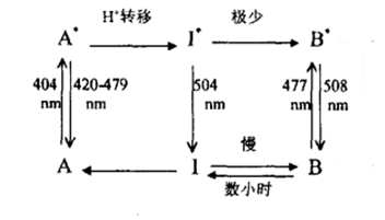 Figure 10. Model of the GFP fluorescence mechanism[12]
 In a minority of cases, changes in the hydrogen-bond network stabilize the chromophore in its ionic form. The His148 side chain forms a hydrogen bond with the Tyr66 phenolic oxygen (Oη), while Wat304 interacts with Tyr66 Oη and the side-chain oxygens of Thr203 and Ser205. At the same time, the hydrogen bond between Glu222 and Ser205 breaks, and the carboxyl group of Glu222 may undergo cis–trans isomerization.
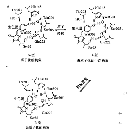 Figure 11. Chromophore conformations in different GFP states[12]
 Engineering Fluorescent Proteins
Wild-type fluorescent proteins have several drawbacks, including weak fluorescence, temperature-sensitive folding, slow maturation, and, in the case of drFP583, cytotoxicity. Most fluorescent proteins used today have therefore been engineered. These modifications usually focus on the chromophore and follow one of two strategies: alter the chromophore itself to change its photochemical properties, or replace amino acids in its immediate environment to change how it fluoresces. Several common engineered proteins are described below[11].
Enhanced Blue Fluorescent Protein (EBFP)
EBFP replaces Tyr66 in wild-type GFP with His, substituting an imidazole group for the original phenolic group. The mutation shifts both spectra toward the blue (excitation: 383 nm; emission: 445 nm). The additional mutations Val150Ile and Val224Arg fill the cavity left by the tyrosine-to-histidine substitution and stabilize the chromophore.
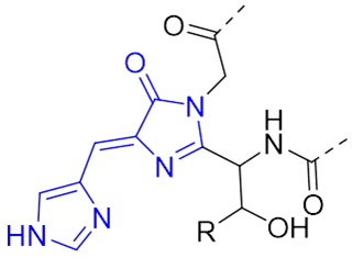 Figure 12. Structure of the EBFP chromophore
 Enhanced Cyan Fluorescent Protein (ECFP)
ECFP follows the same basic strategy as blue fluorescent protein. Replacing Tyr66 with tryptophan introduces an indole group into the chromophore. Its excitation and emission wavelengths then fall between those of the neutral and ionic phenolic forms of wild-type GFP, producing characteristic blue-green fluorescence (Ex: 439 nm; Em: 476 nm). Replacing His148 with aspartic acid also removes the interaction between Tyr145 and His148, stabilizing the protein's first conformational state. A Tyr145Ala substitution further increases fluorescence intensity, while S72A helps the chromophore mature more quickly.
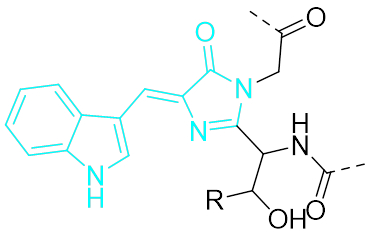 Figure 13. Structure of the ECFP chromophore
 Enhanced Yellow Fluorescent Protein (EYFP)
EYFP was created mainly by engineering the microenvironment around the wild-type GFP chromophore. The most important change from avGFP is the replacement of Thr203 with tyrosine. When the protein folds, the Tyr203 phenolic group extends into the internal cavity and forms a π–π stacking interaction with the chromophore's phenolic group. This mutation stabilizes the chromophore's excited-state dipole moment and shifts both absorption and emission about 20 nm toward the red. The Gln69Lys mutation increases chromophore ionization and moves the emission wavelength another 1–2 nm in the same direction.
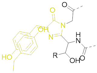 Figure 14. Structure of the EYFP chromophore
 Superfolder GFP
Superfolder GFP combines nine point mutations: S30R, Y39N, F99S, N105T, Y145F, M153T, V163A, I171V, and A260V. It is more stable than wild-type GFP, folds more efficiently even at 37 ℃, and is brighter than EGFP. It also withstands acidic and alkaline conditions better than other members of the GFP family.
Orange Fluorescent Protein (mOrange)
mOrange belongs to the mFruit family and has characteristic absorption and emission wavelengths of 548 nm and 562 nm, respectively. Engineered from the red fluorescent protein DsRed, mFruit proteins are monomeric, bright, quick to mature, and relatively nontoxic. mOrange fluoresces orange because an oxazole ring forms at Thr66 as the chromophore develops. This ring reduces conjugation of the carbonyl group at residue 65, shifting the absorption and emission spectra toward the blue relative to the wild type.
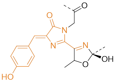 Figure 15. Structure of the mOrange chromophore
 The field has come a long way from the discovery of a single green fluorescent protein. Biologists can now choose among many proteins with different spectral properties, and these versatile labels have transformed research across the life sciences. Continued work on fluorescent proteins will give researchers an even wider range of tools.
Main References
[1] Zhu Yun, Li Chaowei. Green Fluorescent Protein[J]. Biology Teaching, 2010, 35(6): 62.
[2] Wang Yupeng. The Story of Fluorescent Proteins[J]. Biology Teaching, 2019, 44(5): 73-74.
[3] Hao Limei, Li Tangdi. Advances in Red Fluorescent Protein Research[J]. Foreign Medical Sciences: Pharmacy, 2006, 33(2): 131-133.
[4] Liu Mofang, Wang Enduo. Green Fluorescent Protein[J]. Progress in Biochemistry and Biophysics, 2000, 27(3): 238-243.
[5] Liu Zuqiang, Hu Min, Qi Yipeng. The Structure, Fluorescence Mechanism, and Applications of Green Fluorescent Protein[J]. Journal of Wuhan University, 2000, 46(2): 211-214
[6] Remington, S.J., Saif, M. Crystal structure of GFP S205A/T203V at 2.2 A resolution [DB/OL]. www.pdbus.org/structure/4OGS, 2014-05-14/2021-12-20.
[7] Ma Jinshi. Green Fluorescent Protein[J]. Chemistry Bulletin, 2009, 3: 243-250.
[8] Tubbs, J.L., Tainer, J.A., Getzoff, E.D. High Resolution Crystal Structure of the Discosoma Red Fluorescent Protein (DsRed) [DB/OL]. www.pdbus.org/structure/1ZGO, 2005-08-02/2021-12-20.
[9] Fan Jinyu, Cui Zongqiang, Zhang Xian’en. Spectral Diversity and In Vitro Molecular Evolution of Red Fluorescent Proteins[J]. Progress in Biochemistry and Biophysics, 2008, 35(10): 1112-1120.
[10] Head, J.F., Inouye, S., Teranishi, K., Shimomura, O… Crystal Structure of Aequorin[DB/OL]. www.pdbus.org/structure/1EJ3, 2000-05-31/2021-12-21.
[11] Sample V, Newman R H, Zhang J, et al. The structure and function of fluorescent proteins[J]. Chemical Society Reviews, 2009, 38(10): 2852-2864.
[12] Wang Wei, Liu Xianglin, Li Jiudi. The Fluorescence Mechanism of Green Fluorescent Protein[J]. Chemistry of Life, 1999, 19(6): 275-279.

 爱发电(点击图片跳转)