
How the Human Brain Develops: Building the Most Remarkable Object in the Universe
Author: Abaitel
The mature human brain is capable of higher thought and learning, extraordinary creativity and imagination, and a deep appreciation of beauty. It is hard to believe that all of this begins with a single fertilized egg. Drawing on a semester of studying the brain in and outside the classroom, I will sketch a broad but striking picture of how the human brain develops.
Early embryonic development and the fetal brain
In a dramatic long-distance race, one competitor edges ahead of a crowd propelled by beating tails. It reaches the finish line by the narrowest of margins: a warm, spherical cell. This is the egg, the destination of the racing sperm. Each sperm carries different genetic information, but only the one that enters first can combine its information with the egg’s and help shape a new individual.
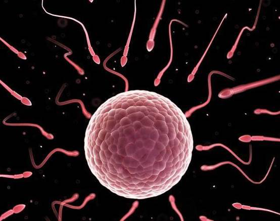
As the successful sperm enters the egg, chemical changes at the egg’s surface block any others from following. The genetic material of the sperm and egg then fuse, forming a zygote. If development proceeds normally, the information in that single cell will eventually reach every part of a new human body.
About 30 hours later, the zygote completes its first division and becomes two cells. The cells keep dividing, and after three days they form a cluster that resembles a mulberry, with each “seed” representing one cell. This stage is aptly named the morula.
Five days after fertilization, the morula undergoes a clear change. Instead of remaining a solid cluster, its cells separate into two groups. One forms an outer wall around the other, the inner cell mass. The result looks a little like a hollow plastic ball with a smaller ball stuck to one side inside it. The morula is now a blastocyst. Its outer layer supports and nourishes the embryo, while the embryo itself develops from the inner cell mass.
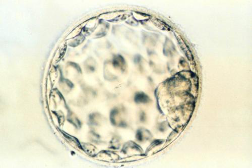
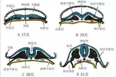
The rounded blastocyst then implants in the lining of the uterus. The inner cell mass pulls away from the outer wall and forms a two-layer embryonic disc. About 12 days after implantation, some cells from the upper layer move between the two existing layers and create a third. Chemical signals from this new layer cause the uppermost cells to begin differentiating into neurons, turning that layer into the neural plate. The neural plate will give rise to the nervous system, including the brain.
Between 18 and 20 days after implantation, the neural plate begins to change shape. During the third week, its two edges rise, creating the neural groove, and then curl inward until they meet above the groove to form the neural tube. The first outline of the brain is now in place. The tube’s cavity later becomes the brain’s ventricular system, where cerebrospinal fluid circulates, carrying nutrients and waste products.
Once the neural tube has formed, neural cells continue dividing and the early brain keeps growing. Two months after implantation, differences in growth rate make separate brain regions recognizable. The paired bulges on either side will become the cerebral hemispheres, while a later projection at the rear will become the cerebellum.
The brain can develop distinct structures only as its population of neural cells grows. Neural progenitor cells repeatedly travel between the outer edge and the center of the neural tube as they divide. A cell preparing to divide moves inward, divides near the center, and sends its daughter cells back toward the periphery to grow before the next round. As this continues, the new cells must acquire specialized roles and reach the right functional regions. They move in different directions and become several kinds of cells. Some become neurons; others become macrophages that clear debris, myelinating cells that provide insulation, or astrocytes that help keep the local environment stable. Neurons may have the starring role, but these glial cells are indispensable too.
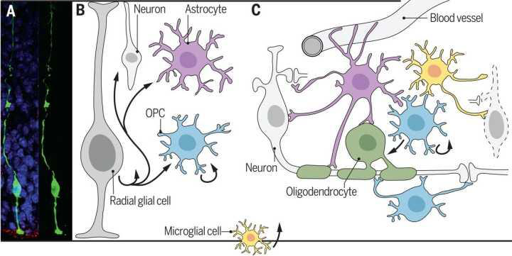
Glial cells lay down tracks that guide neurons from the center toward other parts of the brain. Particular neurons follow particular tracks, moving outward until they reach their destinations and peel away. We still do not know exactly how they leave those tracks or gather into functional groups. What we do know is that the brain grows as newly formed neurons migrate outward and settle into place. Unlike the cortex of many other animals, the human cerebral cortex builds its innermost layer first, then stacks new layers outside it until all six have formed. Parts of the skull remain flexible as the brain expands. Because a flat sheet of cortical cell bodies would require an enormous area, the cortex folds into gyri and sulci, packing more surface area into a smaller volume. Researchers have reproduced this folding with a simple physical model.
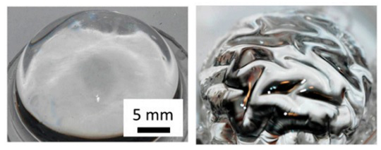
By the ninth month of pregnancy, the fetal brain contains almost as many neurons as an adult brain. At roughly 350 cubic centimeters, it is about the size of a chimpanzee’s brain. Any larger and it could no longer pass easily through the mother’s birth canal, so birth must occur. With the number of neurons already fairly stable, cell division cannot explain how the brain later grows to four times this volume. What drives that growth, and what gives us abilities no newborn could possess? The next section looks at some possible answers.
Brain development in infancy and early childhood
Birth not only frees the infant brain from the size limit imposed by the mother’s birth canal; it also brings the child into a world far richer in sensory input than the womb. The brain’s job is to respond to the outside world with increasing effectiveness and control, and this flood of new stimulation gives it the opportunity to practice. Birth marks a turning point: before it, brain development depends largely on producing more cells; afterward, the organization and performance of those cells matter more.
The first obvious change is myelination. As noted earlier, some neural stem cells become glia, and some glial cells wrap neuronal processes in myelin, insulating the axons. The amount of myelin in the cerebral cortex rises rapidly during the first month after birth. Better insulation lets neural signals travel farther with less loss, a prerequisite for fine motor control. Precise movement requires the cortex to send different electrical signals along neural pathways to distant effectors, and that works only when the signals travel efficiently.
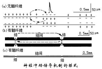
By about one year of age, infants often reach for objects without thinking. At first they can move their fingers only as a group, but as they grow they learn to move each finger separately. This may reflect further specialization among the brain’s functional regions. More complex and precise neural activity depends closely on the growth of neuronal processes and the formation of synapses. Much of the brain’s remarkable increase in volume after birth comes from these intricate connections, which increase the space occupied by each neuron. Neural fibers sometimes grow between neurons far apart from one another. Some researchers think chemicals released by distant neurons attract the fibers; others propose that pioneer fibers formed early in development clear a path for those that follow.
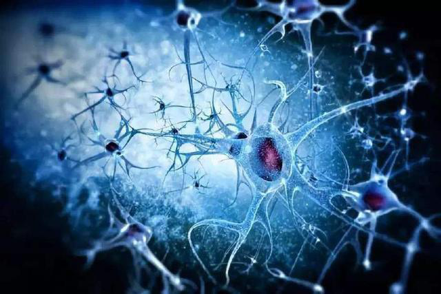
Neural connections are not simple one-to-one pairings fixed in advance; they change in response to the environment. An Italian boy, for example, was found to be blind in one eye even though the eye itself appeared normal. As an infant, he had developed a mild infection in that eye and worn a bandage over it for two weeks. With no input from the covered eye, the brain treated it as unusable, and connections from the other eye claimed cortical territory that would normally serve both. By the time the bandage came off, the affected eye could no longer establish effective connections with its target region, and the cortex could not receive its signals.
As myelination spreads and neural connections multiply through infancy and early childhood, clear signs of consciousness emerge along with a growing ability to solve problems. Does the brain stop developing then? Not at all.
Later brain development
The brain continues developing throughout life. Connections formed early on may later disappear, leaving those best suited to a person’s traits and surroundings. We are shaped by both our genes and our experiences, and as the environment changes, the brain keeps changing in subtle ways.
The brain is generally thought to mature after another 11 years of development beyond age five. Over that period, it grows by another 5 percent. That final 5 percent may help determine what we become good at and what work we choose. Experience continues to fine-tune the brain afterward, although the changes grow smaller and less frequent. The amount of stimulation helps determine how many neural connections form during development, while the type of stimulation selectively activates and strengthens particular circuits. As adults, we should therefore seek out worthwhile experiences and practice, using beneficial stimulation to shape our neural connections and, in turn, ourselves.
As we enter middle and old age, the brain inevitably begins to decline. By age 70, a healthy person’s brain may have lost 5 percent of its earlier weight; by age 90, the loss may reach 20 percent. Still more dangerous are neurodegenerative diseases, which can take away a patient’s abilities, memories, sense of self, and eventually almost everything else. Caring for people with these disorders in an aging world is now one of humanity’s major challenges.
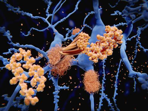
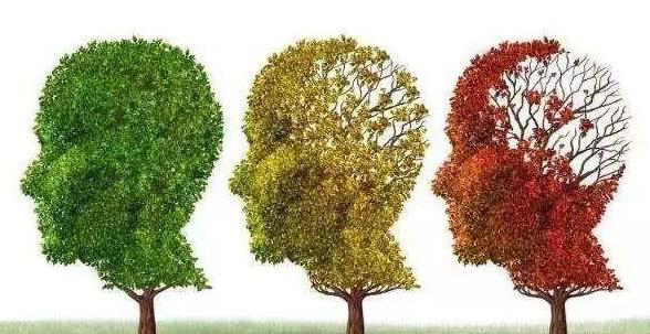
This is the life of the brain as I see it. It is also the life of each one of us. If we want to understand our own lives, learning about the brain and the science behind it is both valuable and essential.
 爱发电(点击图片跳转)